RESPIRATORY
PNEUMOTHORAX (NON TRAUMA)
Prompt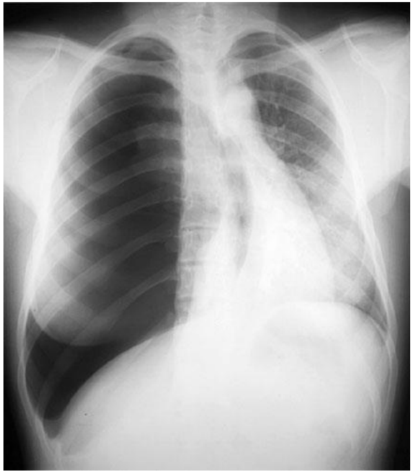
ResponseRight pneumothorax
Significant tension with massive mediastinal shift
Flattened right hemidiaphragm
Small amount right pleural fluid
Deep sulcus
No subcutaneous emphysema
No medical intervention (lines, drains etc)
Trachea midline
No focal consolidation
Significant tension with massive mediastinal shift
Flattened right hemidiaphragm
Small amount right pleural fluid
Deep sulcus
No subcutaneous emphysema
No medical intervention (lines, drains etc)
Trachea midline
No focal consolidation
Prompt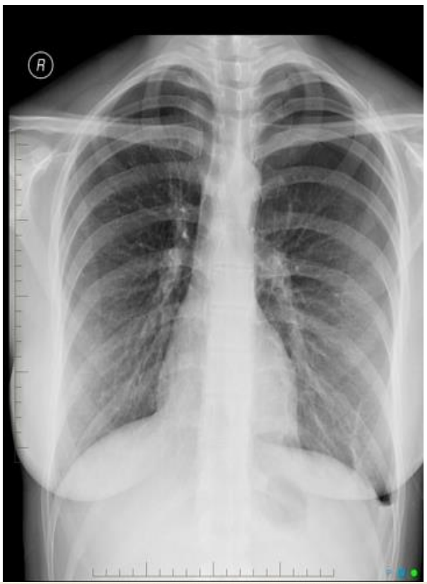
ResponseLeft sided pneumothorax - BST guideline moderate to large - 1-2cm intrerpleural measured at the hilum
Prompt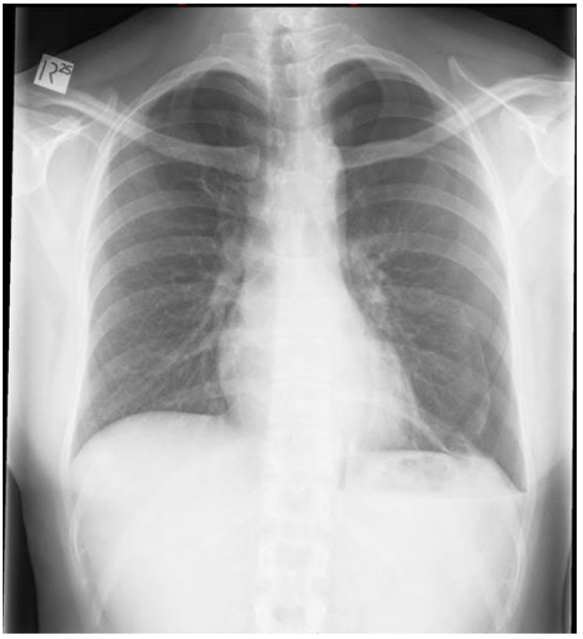
ResponseLeft sided pneumothorax
Subtle pneumomediastinum present (For bonus marks!)
Subtle pneumomediastinum present (For bonus marks!)
Prompt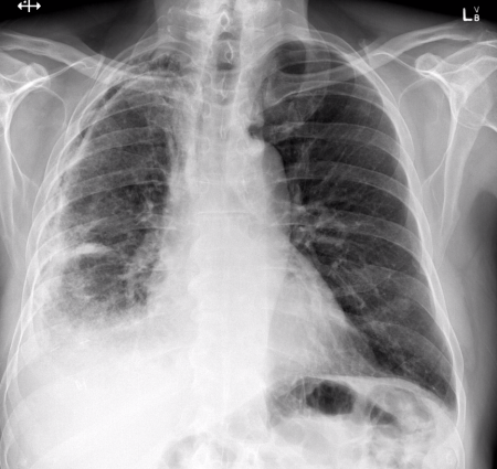
ResponseR pneumothorax - visible pleural line with absent lung markings
Pleural thickening/plaque - broad opacification (widespread) along right visceral pleura
Right sided pleural effusion (consolidation with effusion) - loss of right hemidiaphragm with meniscus sign
Interstitial lung disease - increased interstitial markings right lung
Pleural thickening/plaque - broad opacification (widespread) along right visceral pleura
Right sided pleural effusion (consolidation with effusion) - loss of right hemidiaphragm with meniscus sign
Interstitial lung disease - increased interstitial markings right lung
Prompt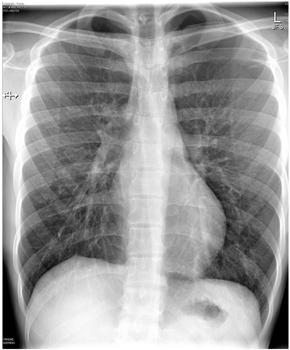
ResponseLeft sided pneumothorax
Visible lung edge
Absence of lung markings at left apex.
Visible lung edge
Absence of lung markings at left apex.
Prompt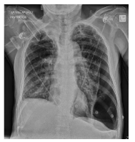
ResponseLeft sided pulmonary bulla
Left sided loculated pneumothorax
Hyperiflated left hemithorax
Emphysematous change or scarring to background lungs
NOn invasive ventilation circuit
?bullae R side upper zone
Mgmt - Immediately stop NIV and change to NRBM
Predicted US findings
R - Lung sliding present, A lines
L - Absence of lung sliding, lung point, barcode sign
Left sided loculated pneumothorax
Hyperiflated left hemithorax
Emphysematous change or scarring to background lungs
NOn invasive ventilation circuit
?bullae R side upper zone
Mgmt - Immediately stop NIV and change to NRBM
Predicted US findings
R - Lung sliding present, A lines
L - Absence of lung sliding, lung point, barcode sign
Prompt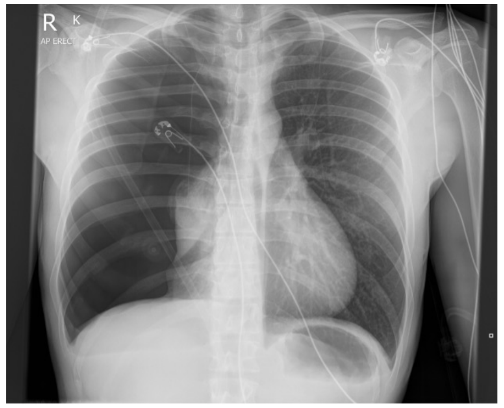
ResponseLarge right sided pneumothorax (~70%)
Shift to left (radiographic tension)
Flattened hemi-diaphragm
Collapse involves all lobes
Pneumomediastinum present
Absence of lung markings
Hemithorax radiolucent compared to left
Shift to left (radiographic tension)
Flattened hemi-diaphragm
Collapse involves all lobes
Pneumomediastinum present
Absence of lung markings
Hemithorax radiolucent compared to left
Prompt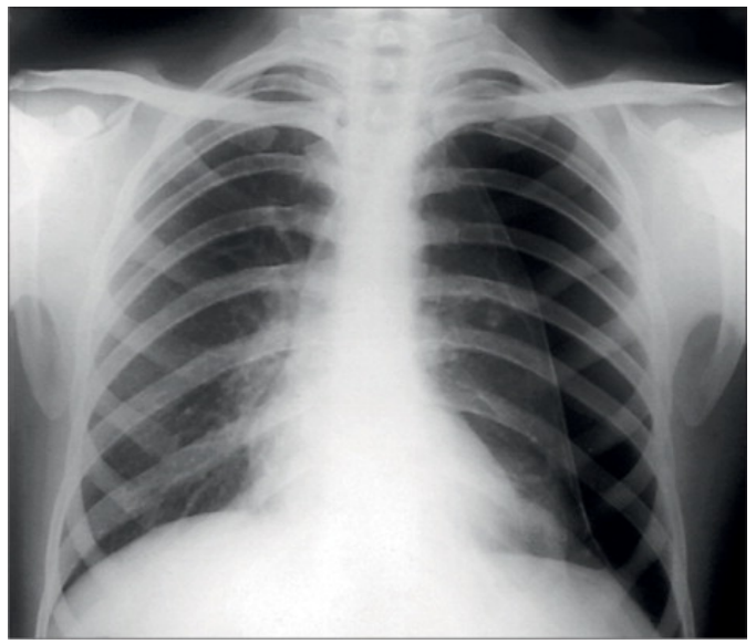
ResponseI give up. You should be able to do this by now
PromptWhats this show?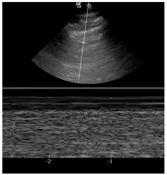
ResponsePleural sliding
PromptWhats this show?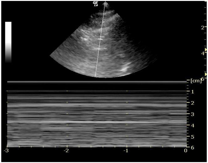
ResponsePneumothorax
Barcode sign = Stratosphere sign
No motion of chest wall
No motion of lung
(opposite = Seashore sign = No ptx)
Barcode sign = Stratosphere sign
No motion of chest wall
No motion of lung
(opposite = Seashore sign = No ptx)
PromptWhats this show?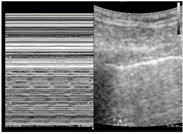
ResponseNo pneumothorax
“Lung pulse” - repeated vertical lines that are seen only below the pleural line. Represents the lungs Jiggle as oxygenated blood fils it. Artifact useful as not seen in PTx, only seen in lung, even unventilated lung.
“Lung pulse” - repeated vertical lines that are seen only below the pleural line. Represents the lungs Jiggle as oxygenated blood fils it. Artifact useful as not seen in PTx, only seen in lung, even unventilated lung.
PromptNormal lung sliding?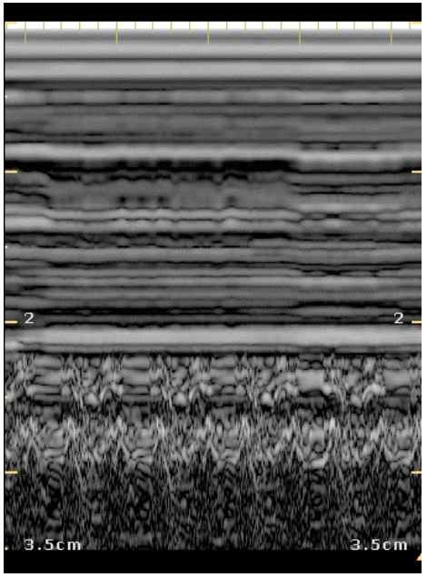
ResponseNo. Trick question
This is an M mode image of cardiac motion just under the chest wall
This is an M mode image of cardiac motion just under the chest wall
- Lung should be bright but this is dark (as filled with blood)
- Zig-zag of movement represents movement toward/away from probe (and the lung doesn’t do this normally… except in effusions, but its with respiration not heartbeat)
PromptIs this a tension?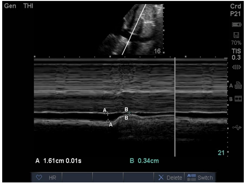
ResponseImage shows IVC that collapses almost completely on inspiration. Intrathoracic pressure is very low on inspiration - therefore not a tension
PromptIs this a tension?
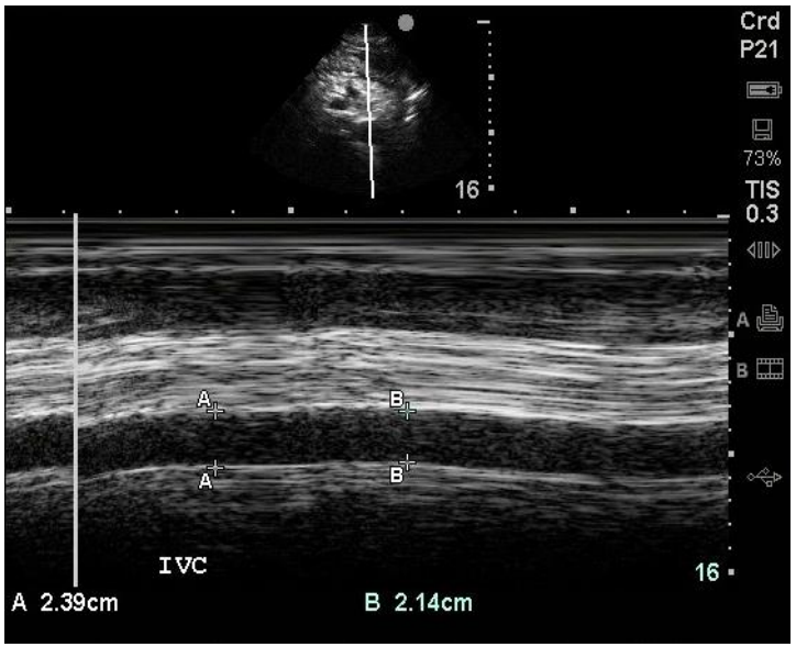
ResponseIVC is behaving like a “Lead pipe” and unchanged - Suggesting high intrathoracic pressure and tension ptx
PromptUS findings suggestive of pneumothorax
ResponseAbsence of lung sliding
Absence of comet tail artifacts
Presence of lung point sign
Loss of normal granular pattern of lung in M mode (absence of seashore sign, presence of barcode/stratosphere sign)
Lead pipe IVC suggests Tension
Absence of comet tail artifacts
Presence of lung point sign
Loss of normal granular pattern of lung in M mode (absence of seashore sign, presence of barcode/stratosphere sign)
Lead pipe IVC suggests Tension
PromptCauses of pneumothorax
ResponsePrimary spontaneous (Smoking is biggest risk factor!)
Secondary spontaneous (Increased mortality)
M:F 6:1
Secondary spontaneous (Increased mortality)
- Asthma, COPD, TB, pneumonia, lung abscess, carcinoma, CF, Fibrosis
- Inhallation/insufflation/snorting/IVDU
- Collagen vascular disease (eg: Marfan’s syndrome)
M:F 6:1
PromptImportant historic features of pneumothorax
ResponsePrimary vs secondary - Secondary/recurrent more likely to require intervention
Prior pneumothorax/ipsilateral pneumothorax
Known lung disease
Symptomatology
breathless/chest pain - more symptomatic = more likely tor equire intervention
Patient factors
Consent/wishes/special considerations (Employment - diver/pilot etc)
Prior pneumothorax/ipsilateral pneumothorax
Known lung disease
Symptomatology
breathless/chest pain - more symptomatic = more likely tor equire intervention
Patient factors
Consent/wishes/special considerations (Employment - diver/pilot etc)
PromptClinical features of tension pneumothorax
ResponseHypoxia
Hypotension
Agitation
Dyspnoea
Shock
Ipsilateral dullness to percussion
Ipsilateral decreased air entry
Contralateral hyperexpansion appearance
(Tracheal deviation is not a reliable sign)
Hypotension
Agitation
Dyspnoea
Shock
Ipsilateral dullness to percussion
Ipsilateral decreased air entry
Contralateral hyperexpansion appearance
(Tracheal deviation is not a reliable sign)
PromptRadiologic features that may influence management
ResponseLarge vs small
Midline shift/radiologic tension
BTS vs ATS rules - eg: >2cm interpleural = large - more likely to require intervention
Midline shift/radiologic tension
BTS vs ATS rules - eg: >2cm interpleural = large - more likely to require intervention
PromptPre-hospital ultrasound to confirm pneumothorax - pros and cons
ResponsePros - Fast, no radiation, portible, accurate with training (High specificicity/sensitivity), potentially avoids unnecessary intervention
Cons - Requires training, ambient bright light may make interpretation difficult, patient access may be challenging, s/c emphysema may cause artefact
Cons - Requires training, ambient bright light may make interpretation difficult, patient access may be challenging, s/c emphysema may cause artefact
PromptNeedle decompression - summarise pros and cons
ResponseRapid, can be done whilst patient trapped
Lower level of training required to perform
Patient doesn’t required sedation for procedure
Temporising only and may be ineffective
Landmark accuracy prone to error
Can cause iatrogenic vascular injury and bleeding
Lower level of training required to perform
Patient doesn’t required sedation for procedure
Temporising only and may be ineffective
Landmark accuracy prone to error
Can cause iatrogenic vascular injury and bleeding
PromptFinger thoracostomy - summarise pros and cons
ResponseNeed 360 degree access, slower than needle decompression
Definitive management
Landmarks prone to error but larger area of safety vs needle
Iatrogenic intercostal vessel/nerve injury and bleeding
Needs more training to perform
Needs local anaesthetic +/- sedation
Ideally needs positive pressure ventilation
Definitive management
Landmarks prone to error but larger area of safety vs needle
Iatrogenic intercostal vessel/nerve injury and bleeding
Needs more training to perform
Needs local anaesthetic +/- sedation
Ideally needs positive pressure ventilation
PromptIndications for chest drain without imaging in suspected pneumothorax
ResponseSaturations <92% on oxygen
SBP <90
RR <10
Decreased LOC on oxygen
Pre-arrest/cardiac arrest
SBP <90
RR <10
Decreased LOC on oxygen
Pre-arrest/cardiac arrest
PromptImmediate management of tension pneumothorax
ResponseImmediate chest decompression - Needle thoracocentesis, ICC if rapid, Finger thoracostomy then 3 sided occlusive dressing
Ensure improvement of clinical state and vital signs
Non invasive monitoring
Formal ICC with underwater seal drain
Oxygen
Manage underlying condition (Eg: Asthma give nebs)
Ensure improvement of clinical state and vital signs
Non invasive monitoring
Formal ICC with underwater seal drain
Oxygen
Manage underlying condition (Eg: Asthma give nebs)
PromptManagement options for pneumothorax
ResponseConservative
Minimal symptomatology at rest, first primary pneumothorax wsuspected without obvious underlying structural abnormality on CXR. Stable patient on observation.Patient preference. Get progress film in 6-12 hours for stability, follow up film 2 weeks
Apparently 94% success rate for spontaneous Ptx.
Simple aspiration
16-18 ga needle, <2.5L. Faster resolution of symptoms/size in spontaneous pneumothroax. Patient preference, promotes earlier discharge
Chest drain
Pigtail 8-14F. Secondary pneumothorax suspected (recurrence/underlying lung disease), significant symptoms - chest discomfort not controlled with simple analgesia
ED indications:
Progressive hypoxia despite supplemental O2
Worsening fatigue (resp acidosis)
Progressive/increasing ptx on serial XR
Primary VATS/Pleurodesis
Access to appropriate specialty
Work related requirement (Eg: Pilot)
Recurrent Ptx
Minimal symptomatology at rest, first primary pneumothorax wsuspected without obvious underlying structural abnormality on CXR. Stable patient on observation.Patient preference. Get progress film in 6-12 hours for stability, follow up film 2 weeks
Apparently 94% success rate for spontaneous Ptx.
Simple aspiration
16-18 ga needle, <2.5L. Faster resolution of symptoms/size in spontaneous pneumothroax. Patient preference, promotes earlier discharge
Chest drain
Pigtail 8-14F. Secondary pneumothorax suspected (recurrence/underlying lung disease), significant symptoms - chest discomfort not controlled with simple analgesia
ED indications:
Progressive hypoxia despite supplemental O2
Worsening fatigue (resp acidosis)
Progressive/increasing ptx on serial XR
Primary VATS/Pleurodesis
Access to appropriate specialty
Work related requirement (Eg: Pilot)
Recurrent Ptx
PromptLandmarks for drainage
ResponseTriangle of safety:
Anterior Border: Lateral edge of the pectoralis major muscle.
Posterior Border: Lateral edge of the latissimus dorsi muscle.
Inferior Border: A horizontal line usually corresponding to the 4th or 5th intercostal space (just above the nipple line).
Apex: The armpit (axilla)
Anterior Border: Lateral edge of the pectoralis major muscle.
Posterior Border: Lateral edge of the latissimus dorsi muscle.
Inferior Border: A horizontal line usually corresponding to the 4th or 5th intercostal space (just above the nipple line).
Apex: The armpit (axilla)
PromptContraindications for ED ICC
ResponsePatient refusal
Iatrogenic coagulopathy (DOAC/Warfarin)
Marked thrombocytopaenia
Active malignancy with risk of dissemination
Established ceiling of care stating not for invasive measures
Iatrogenic coagulopathy (DOAC/Warfarin)
Marked thrombocytopaenia
Active malignancy with risk of dissemination
Established ceiling of care stating not for invasive measures
PromptComplications of ICC placement
ResponseMisplacement of ICC
Solid organ injury
Infection/empyema
Haemorrhage
Tube malfunction/blockage/kinking
Solid organ injury
Infection/empyema
Haemorrhage
Tube malfunction/blockage/kinking
PromptDischarge advice for pneumothorax
ResponseFollow up organised (LMO vs Resp) + timing
Progress films in 2-4 weeks
High risk activity advice - No flying until full resolution + cleared by resp physician. No diving at any point (Unless sutical fixation and normal CT)
Return advice - worsening CP/SOB
Cessation of smoking
Progress films in 2-4 weeks
High risk activity advice - No flying until full resolution + cleared by resp physician. No diving at any point (Unless sutical fixation and normal CT)
Return advice - worsening CP/SOB
Cessation of smoking