RESPIRATORY
LUNG INFECTION - PNEUMONIA/ABSCESS
All seem to be cavitation lesions
PromptLots of chest Xrays and differentials:
Response
Prompt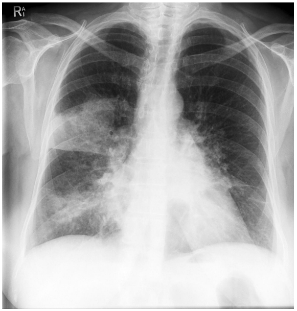
ResponseLeft Hilar mass - round opacity at the left hilum with some patchy consolidation around this area
Confluent consolidation right upper lobe bordered by horizontal fissure
Air bronchograms
Patchy consolidation right lower zone as well - right middle lobe as part of right cardiac border obscured
Dx:
Multi lobar pneumonia
Left hilar mass likely malignancy
Confluent consolidation right upper lobe bordered by horizontal fissure
Air bronchograms
Patchy consolidation right lower zone as well - right middle lobe as part of right cardiac border obscured
Dx:
Multi lobar pneumonia
Left hilar mass likely malignancy
Prompt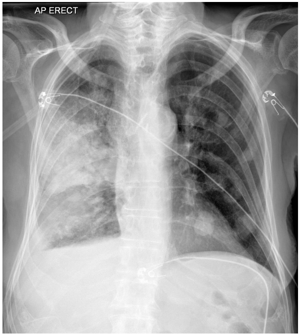
ResponseMultiple round opacities Left lung
Los of volume right hemithorax (Consistent with collapse)
Right upper lobe consolidation
Air bronchograms
R lower lobe opacification/consolidation
Pleural effusion R side (Parapneumonic)
Hilar mass (probably lymphadenopathy R side)
Provisional diagnosis
Multi lobar pneumonia
Malignancy probably metastatic
Los of volume right hemithorax (Consistent with collapse)
Right upper lobe consolidation
Air bronchograms
R lower lobe opacification/consolidation
Pleural effusion R side (Parapneumonic)
Hilar mass (probably lymphadenopathy R side)
Provisional diagnosis
Multi lobar pneumonia
Malignancy probably metastatic
Prompt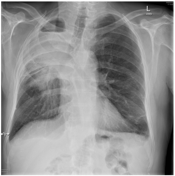
ResponseRUL opacification
Cavitation lesion with air fluid level
RUL collapse - tracheal deviation and R hemithorax volume loss with elevation of horizontal fissure
Opacification of superior medial segments of RML consistent with mass or consolidation
Small left basal effusion
DDx
Neoplastic - Primary lung (bronchial carcinoma, small cell, non small cell, carcinoid) vs Secondary mets
Lung abscess - bacterial infection - klebsiella, pneumococcus, staph. TB,. other granulmoatous disease
Vasculitis eg: Wegeners
Cavitation lesion with air fluid level
RUL collapse - tracheal deviation and R hemithorax volume loss with elevation of horizontal fissure
Opacification of superior medial segments of RML consistent with mass or consolidation
Small left basal effusion
DDx
Neoplastic - Primary lung (bronchial carcinoma, small cell, non small cell, carcinoid) vs Secondary mets
Lung abscess - bacterial infection - klebsiella, pneumococcus, staph. TB,. other granulmoatous disease
Vasculitis eg: Wegeners
Prompt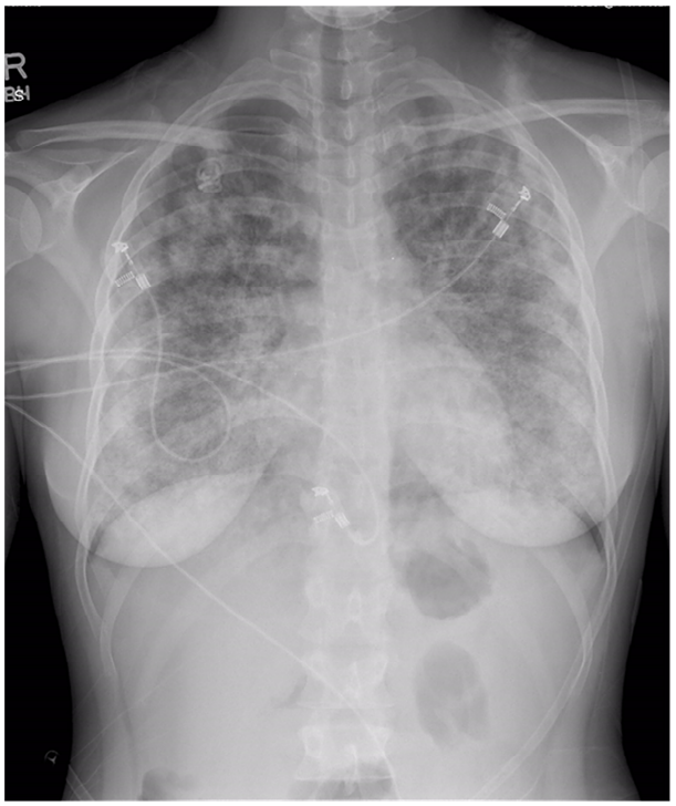
ResponseBilateral symoetrical infiltrates
Alveolar infiltarates with some confluent/round lesions
No effusions
Normal heart size
DDx
Infectious:
Mycoplasma pneumonia
TB
Aspergillus
Influenza
Non infectious
Pulmonary haemorrrhage eg: Wegeners
Lymphangitis carcinomatosis
Pulmonary oedema
Leukaemia
Alveolar proteinosis
Alveolar infiltarates with some confluent/round lesions
No effusions
Normal heart size
DDx
Infectious:
Mycoplasma pneumonia
TB
Aspergillus
Influenza
Non infectious
Pulmonary haemorrrhage eg: Wegeners
Lymphangitis carcinomatosis
Pulmonary oedema
Leukaemia
Alveolar proteinosis
Prompt57M, homeless, EtOH and unkempt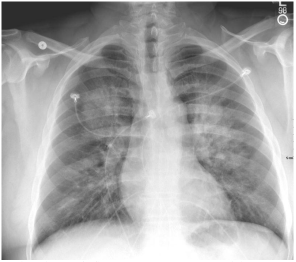
ResponsePatchy alveolar opacification
Predominantly in bilateral perihilar upper lobes
No hilar lymphadenopathy
Cardiac silhouettes normal, normal cardiothoracic ratio
No effusion
No rib fractures or ptx
DDx
Alveolar haemorrhage (underlying lung disease, vasculitis, coagulopathy or drug/cocaine abuse)
Acute pulmonary oedema (Cardiogenic vs non cardiogenic)
Infection
Septic emboli from cardiac valvular disease/IE
Predominantly in bilateral perihilar upper lobes
No hilar lymphadenopathy
Cardiac silhouettes normal, normal cardiothoracic ratio
No effusion
No rib fractures or ptx
DDx
Alveolar haemorrhage (underlying lung disease, vasculitis, coagulopathy or drug/cocaine abuse)
Acute pulmonary oedema (Cardiogenic vs non cardiogenic)
Infection
- bacterial ?Staph, viral or atypical pneumonia
- Immunocompromised ?PCP/TB
- Aspiration due to obtundation/alcohol use
Septic emboli from cardiac valvular disease/IE
Prompt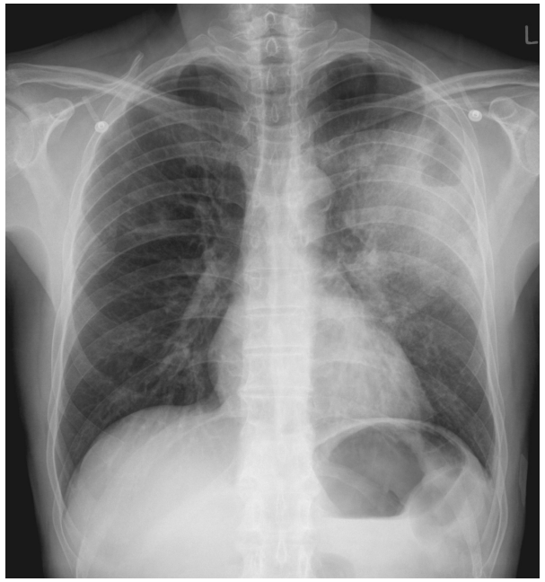
ResponseLeft upper zone cavitation lesion
Fluid level
Surrounding left upper zone consolidation
Air bronchograms
No mediastinal lymphadenopathy
Solitary area of pathology
No effusion
DDx
S. Pneumonia
S. Aureus
Klebsiella
TB
Fungal - Cryptococcus
Non Infectious DDx
Malignancy
Granuloma (eg: Wegeners)
Infarct
Bronchogenic cyst
Fluid level
Surrounding left upper zone consolidation
Air bronchograms
No mediastinal lymphadenopathy
Solitary area of pathology
No effusion
DDx
S. Pneumonia
S. Aureus
Klebsiella
TB
Fungal - Cryptococcus
Non Infectious DDx
Malignancy
Granuloma (eg: Wegeners)
Infarct
Bronchogenic cyst
Prompt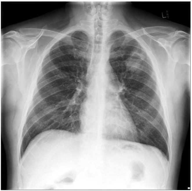
ResponseSolitary lesion Left upper zone
round/oval shaped
Defined wall/discrete lesion
Contains air/fluid level
Cavitating lesion
No surrounding consolidation
No evidence of chronic lung disease
No hilar lymphadenopathy
No PTx
DDx
infection/abscess - Bacterial (Stapch, strep, Klebsieally), TB, fungal
Malignant - primary bronchogenic, metastasis
Granuloma- rheumatoid, wegeners
Infarction - trauma/PE
round/oval shaped
Defined wall/discrete lesion
Contains air/fluid level
Cavitating lesion
No surrounding consolidation
No evidence of chronic lung disease
No hilar lymphadenopathy
No PTx
DDx
infection/abscess - Bacterial (Stapch, strep, Klebsieally), TB, fungal
Malignant - primary bronchogenic, metastasis
Granuloma- rheumatoid, wegeners
Infarction - trauma/PE
Prompt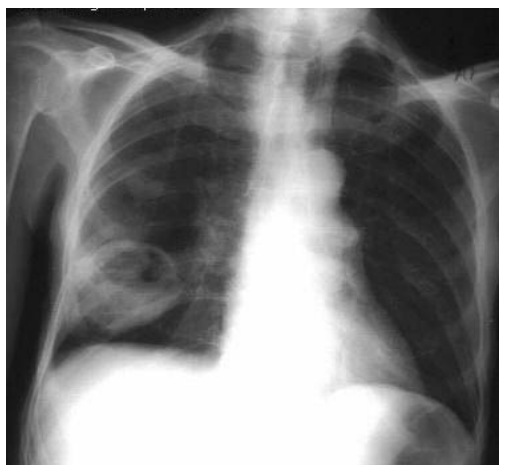
ResponseWell circumscribed thick walled lesion with radiolucent centre in lower lobe of right lung
Consistent with cavitation lesion
Most likely Dx is Lung abscess
DDx
Infectious - Klebsiella, gram neg bacilli, s. Aureus, TB, fungal
Carcinoma - Primary lung, secondary mets
Infarction (from PE), AVM
Consistent with cavitation lesion
Most likely Dx is Lung abscess
DDx
Infectious - Klebsiella, gram neg bacilli, s. Aureus, TB, fungal
Carcinoma - Primary lung, secondary mets
Infarction (from PE), AVM
Prompt86 acute dyspnoea b/g smoker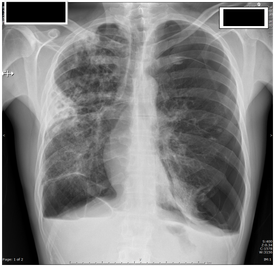
ResponseMarked hyperinflation
Moderate/large L sided pneumothorax
Mediastinal shift to R - possible tension
Bilateral widespread pulmonary infiltrates
Right midzone (Basal segment of upper lobe) wedge shaped cavitation lesion/consoildation
Right apical focal consolidation
Interposed hepatic flexure under right hemidiaphgragm (Chilaiditti syndrome)
DDx
Cavitating pneumonia (Klebisiella, S. Aureus, Pneumococcus, TB)
Malignancy (primary bronchogenic CCA
Intersitial pneumonitis - viral (flu)
Pneumothorax - rupture of large bullae
Moderate/large L sided pneumothorax
Mediastinal shift to R - possible tension
Bilateral widespread pulmonary infiltrates
Right midzone (Basal segment of upper lobe) wedge shaped cavitation lesion/consoildation
Right apical focal consolidation
Interposed hepatic flexure under right hemidiaphgragm (Chilaiditti syndrome)
DDx
Cavitating pneumonia (Klebisiella, S. Aureus, Pneumococcus, TB)
Malignancy (primary bronchogenic CCA
Intersitial pneumonitis - viral (flu)
Pneumothorax - rupture of large bullae
PromptInvestigation to clarify CXR changes
ResponseSputum cultures - ? bacterial causes, Sputum microscopy ? malignant cells
Sputum PCR (Strep/staph)
TB stains - smears on sputum and cultures - positive smear indicates infectivity, culture excludes disease (But takes 4-6 weeks)
Viral PCR (Swabs/NPA) - Conrifm/exclude viral pathogens
CT chest - Support specific diagnosis based on appearance, indicate complications (mets), and can indicate suitability for invasive tests (eg: transbronchial biopsy)
Blood cultures - proved sensitivity for directing antimicrobial therapy in bacterial infection
Echo - LVEF/cardiomyopathy/valve function, ? IE
ECG - Ischaemia as cause for CCF
Vasculitic screen - Goodpastures/Wegners (now Granulomatosis with Polyangitis cos wegner was a Nazi), SLE/RH factor/ANCA/ANA
Sputum PCR (Strep/staph)
TB stains - smears on sputum and cultures - positive smear indicates infectivity, culture excludes disease (But takes 4-6 weeks)
Viral PCR (Swabs/NPA) - Conrifm/exclude viral pathogens
CT chest - Support specific diagnosis based on appearance, indicate complications (mets), and can indicate suitability for invasive tests (eg: transbronchial biopsy)
Blood cultures - proved sensitivity for directing antimicrobial therapy in bacterial infection
Echo - LVEF/cardiomyopathy/valve function, ? IE
ECG - Ischaemia as cause for CCF
Vasculitic screen - Goodpastures/Wegners (now Granulomatosis with Polyangitis cos wegner was a Nazi), SLE/RH factor/ANCA/ANA
PromptManagement of pneumonia
ResponseOxygen - titrate to sats >92%
IV fluids - 500-1000 mL N/S aiming HR <100, SBP >100
IVABx for severe CAP
IV fluids - 500-1000 mL N/S aiming HR <100, SBP >100
IVABx for severe CAP
- IV ceftriaxone 2g daily PLUS azithromycin 500 mg daily
- Moxifloxacin 400 mg IV daily (if penicillin allergy)
- Vancomycin if staph suspected (note says staph not MRSA) - 25-30 mg/kg loading
- Pseudamonas colonised = Tazocin OR Cefepime PLUS Azithro PLUS gent
- eTG ‘24
PromptAntibiotic choice for Lung ABSCESS
Responselung abscess due to aspiration of oral bacteria
Nonsevere
Oral therapy - Amoxicillin 1g TDS + metronidazole 400 mg BD
Clindamycin 450 mg TDS if penicillin allergy
Systemic features or chest wall pain
Benpen 1.2g QID PLUS Metronidazole 400 mg BD PO
Clindamycin 450 mg TDS (PO/IV) if penicillin allergy
Severe disease
Amoxiclav 1g+200mg QID
OR Ceftriaxone 2g OD PLUS Metronidazole 500 mg IV BD
If septic shock add Gent PLUS Vanc
Meropenem 1g TDS if penicillin allergy (+Vanc if shocked)
eTG sept ‘24
Nonsevere
Oral therapy - Amoxicillin 1g TDS + metronidazole 400 mg BD
Clindamycin 450 mg TDS if penicillin allergy
Systemic features or chest wall pain
Benpen 1.2g QID PLUS Metronidazole 400 mg BD PO
Clindamycin 450 mg TDS (PO/IV) if penicillin allergy
Severe disease
Amoxiclav 1g+200mg QID
OR Ceftriaxone 2g OD PLUS Metronidazole 500 mg IV BD
If septic shock add Gent PLUS Vanc
Meropenem 1g TDS if penicillin allergy (+Vanc if shocked)
eTG sept ‘24